

PURCHASED EQUIPMENT
Purchase of an FT-iR (Fourier Transform Infrared) spectroscope, equipped with a microscope and ATR (Attenuated Total Reflectance) module
The acquisition of this equipment develops a new direction of research in NRDI GeoEcoMar, the identification of different types of micropollutants, as well as their quality and quantity, these elements being mandatory for the monitoring of aquatic environments - fresh, brackish and marine.
The abundance of microplastic particles has been steadily increasing since the 1970s, with the introduction of plastic packaging for consumer goods. This type of pollution is the most common and has been observed in all terrestrial and marine natural environments, pollution with a strong impact on all species in the food chain, including humans. For the complete investigation of this type of pollution, in addition to quantitative analyses, it is also necessary to evaluate the types of plastic polymers.
Purchase of an FT-iR (Fourier Transform Infrared) spectroscope, equipped with a microscope and ATR (Attenuated Total Reflectance) module, enables the realization at NRDI GeoEcoMar of the first laboratory in Romania dedicated to microplastic analysis with equipment and instruments for the quantification of microplastics from all types of samples (water, sediment and fauna). The acquisition of an FT-iR spectroscope raises the level of research in this field to the international one, and the scientific results can be capitalized by publication in internationally indexed journals, but also by offering new research services.
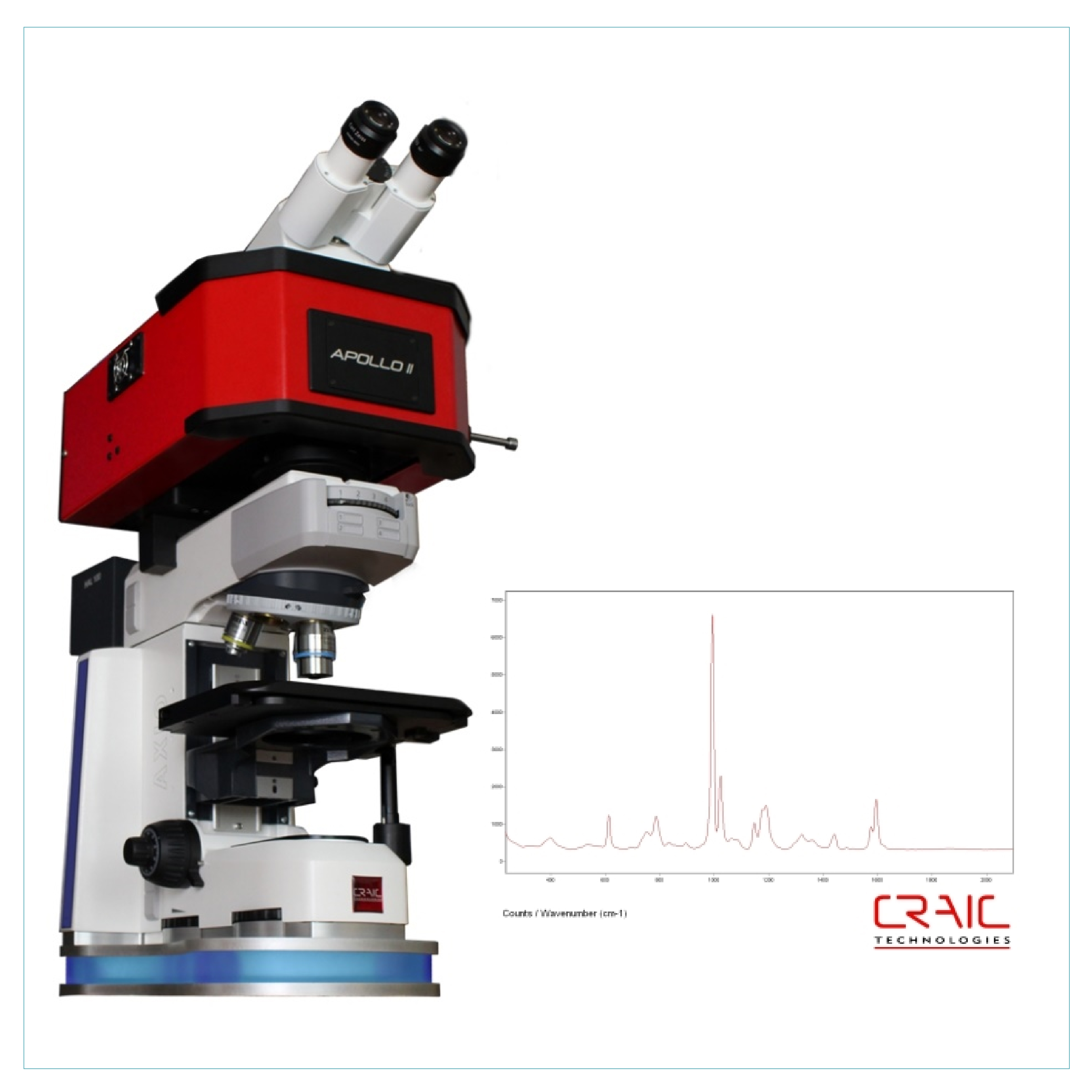
Raman spectroscopy is a spectroscopic technique frequently used in various fields to provide a structural fingerprint by which molecules can be identified. In the case of studies on microplastic particles, RAMAN spectroscopy is used to identify the polymers of the particles, with the aim of evaluating the sources and provenance of these types of waste.
The RAMAN technique is based on inelastic scattering, or Raman scattering of monochromatic light, usually coming from a laser that emits in the visible, near-infrared, or ultraviolet range. The laser light interacts with molecular vibrations, photons, or other excitations in the system, resulting in the energy of the laser photons changing up or down. The energy change provides information about the mode of vibration in the system. Infrared spectroscopy provides similar but complementary information.
The Hardware-Software system of the RAMAN Spectroscope offers the possibility of characterizing microplastic particles according to morphology and polymer type up to 8 µm in size, thus facilitating the visual characterization process performed by a human operator.
SEM (Scanning Electrone Microscope)
A scanning electron microscope (SEM) was purchased as a tool for biology, micropaleontology, chemistry and mineralogy for the study of old and new marine, river and delta systems and, in general, for the study of geology, including marine geology and geo-ecology. The purchased scanning electron microscope (SEM) is used on the full spectrum of microfossils and has a very good field depth; thus, it facilitates the image procurement of the entire analysed space.
The scanning electron microscope (SEM) allows 50000x magnification. This research equipment is the most utilized in the institutions and universities laboratories and has the clearest and largest depth of field, facilitating the image procurement of the entire analysed space. The direct observation allows determining the morphology of the analysed surfaces in the highest of detail. The same quality can be obtained studying minerals from marine sediments.
This research equipment’s acquisition augments the quality of the analyses made on marine, fluvial, coastal and/or deltaic sediments, by approaching an interpretation based on very high-resolution images. By the use of this equipment, the institution will align to the international research requirements.
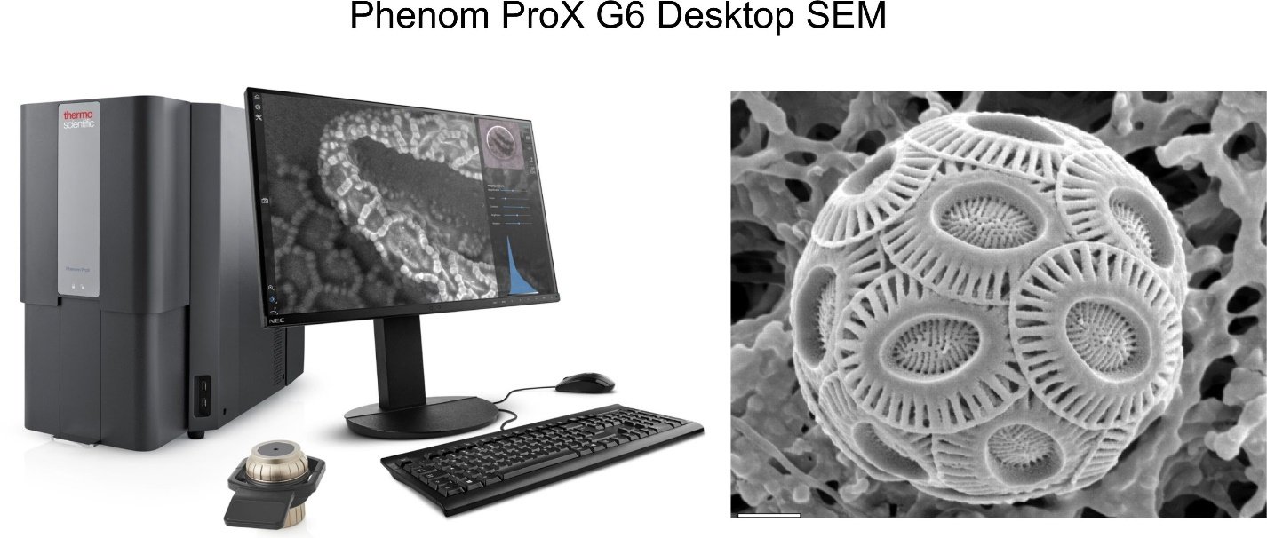
The purchased SEM (Scanning Electron Microscope – left) and an image (right) obtained by using this equipment (calcareous algae from Black Sea, micronic size).